Although meningiomas are the most common primary non-glial intracranial tumors, cystic meningiomas are quite rare. This study presents six cases in order to discuss the radiological and pathological features of cystic meningiomas.
Patients and methodsSix patients with cystic meningiomas were included in the study. All patients underwent a cranial computed tomography scan and magnetic resonance imaging (MRI) evaluation, pre- and postoperatively.
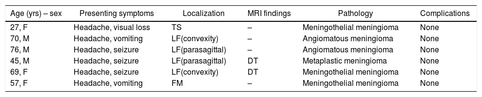
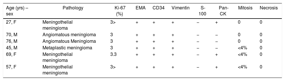
ResultsAll patients presented with long standing headache dating back at least two years. There was no gender predominance in our series. Radiological evaluation revealed two parasagittal and two convexity meningiomas located at the frontal region. Two lesions were located at the tuberculum sellae and the foramen magnum. All of the tumors were totally excised (Simpson Grade I or II). Pathology results included meningothelial meningioma in three patients, angiomatous meningioma in two patients, and metaplastic meningioma in one patient. In two patients, the cystic meningiomas were resected with the use of sodium fluorescein (Na-Fl) under a YELLOW 560nm microscope filter. Na-Fl was found to be very useful in demonstrating the brain–tumor interface, and it was especially effective in resecting the cyst wall of the peritumoural cystic meningiomas. None of the patients had any complications, and no recurrences were noted in any of the patients within the mean follow-up period of 51 months (range: 16–102 months).
ConclusionIt is important to note MRI changes specific to cystic meningioma and include meningiomas in the differential diagnosis of intracranial cystic lesions. The use of sodium fluorescein (Na-Fl) under a YELLOW 560nm microscope filter is a useful tool to differentiate the brain-tumor interface, as well as to identify the cyst wall in order to fully resect the tumor with the cystic component to avoid recurrence and achieve better clinical results.
Aunque los meningiomas son los tumores intracraneales primarios no gliales más frecuentes, los meningiomas quísticos son bastante raros. Este estudio presenta 6 casos para discutir las características radiológicas y patológicas de los meningiomas quísticos.
Pacientes y métodosSe incluyeron 6 pacientes con meningiomas quísticos en el estudio. Todos los pacientes se sometieron a una tomografía computarizada craneal y a una evaluación por resonancia magnética, antes y después de la operación.
ResultadosTodos los pacientes presentaron dolor de cabeza de larga duración de al menos 2 años. No hubo predominio de género. La evaluación radiológica reveló 2 meningiomas parasagitales y 2 de la convexidad ubicados en la región frontal. En los otros 2 pacientes las lesiones se ubicaron en el tubérculo selar y en el foramen magno respectivamente. Todos los tumores fueron totalmente extirpados (grado de Simpson I o II). En 2 pacientes, los meningiomas quísticos se resecaron con el uso de fluoresceína de sodio bajo un filtro de microscopio AMARILLO de 560nm. Se encontró que la fluoresceína de sodio era muy útil para demostrar la interfaz entre el cerebro y el tumor, y fue especialmente eficaz para resecar la pared de los quistes peritumorales. Los resultados de histopatología incluyeron meningioma meningotelial en 3 pacientes, meningioma angiomatoso en 2 y meningioma metaplásico en uno. Ninguno de los pacientes presentó ninguna complicación, y no se observaron recurrencias en ningún paciente dentro del período de seguimiento medio de 51 meses (rango: 16-102 meses).
ConclusiónEs importante tener en cuenta los cambios en la resonancia magnética específicos de los meningiomas quísticos e incluir estos tumores en el diagnóstico diferencial de las lesiones quísticas intracraneales. El uso de fluoresceína de sodio bajo un filtro de microscopio AMARILLO de 560nm es una herramienta útil para diferenciar la interfaz cerebro-tumor, así como para identificar la pared del quiste y resecar completamente el tumor con el componente quístico para evitar la recidiva y conseguir mejores resultados clínicos.
Artículo
![]()
Si es la primera vez que accede a la web puede obtener sus claves de acceso poniéndose en contacto con Elsevier España en suscripciones@elsevier.com o a través de su teléfono de Atención al Cliente 902 88 87 40 si llama desde territorio español o del +34 932 418 800 (de 9 a 18h., GMT + 1) si lo hace desde el extranjero.
Si ya tiene sus datos de acceso, clique aquí.
Si olvidó su clave de acceso puede recuperarla clicando aquí y seleccionando la opción "He olvidado mi contraseña".