


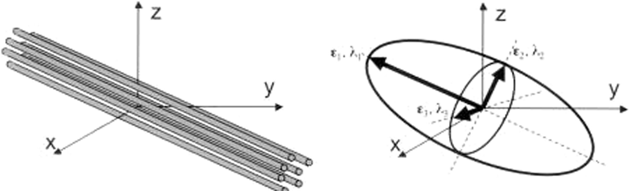
La lesión axonal traumática (LAT) contribuye significativamente a la mortalidad y morbilidad tras traumatismo craneoencefálico (TCE). Sin embargo, la caracterización de la LAT supone un reto diagnóstico para las técnicas de neuroimagen habitual. La secuencia de RM tensor de difusión (diffusion tensor imaging [DTI]) es capaz de detectar el grado de difusión de las moléculas de agua tisular y así inferir la afectación traumática de la sustancia blanca. El objetivo principal de este trabajo ha sido caracterizar la LAT a través de la secuencia DTI realizada en la fase subaguda precoz en nuestra serie de pacientes con TCE moderado y grave y evaluar si existe asociación con la evolución de los pacientes.
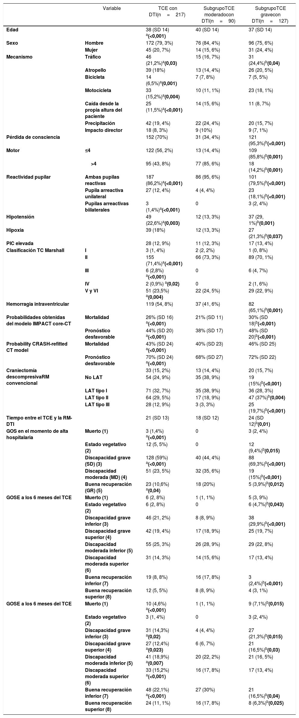
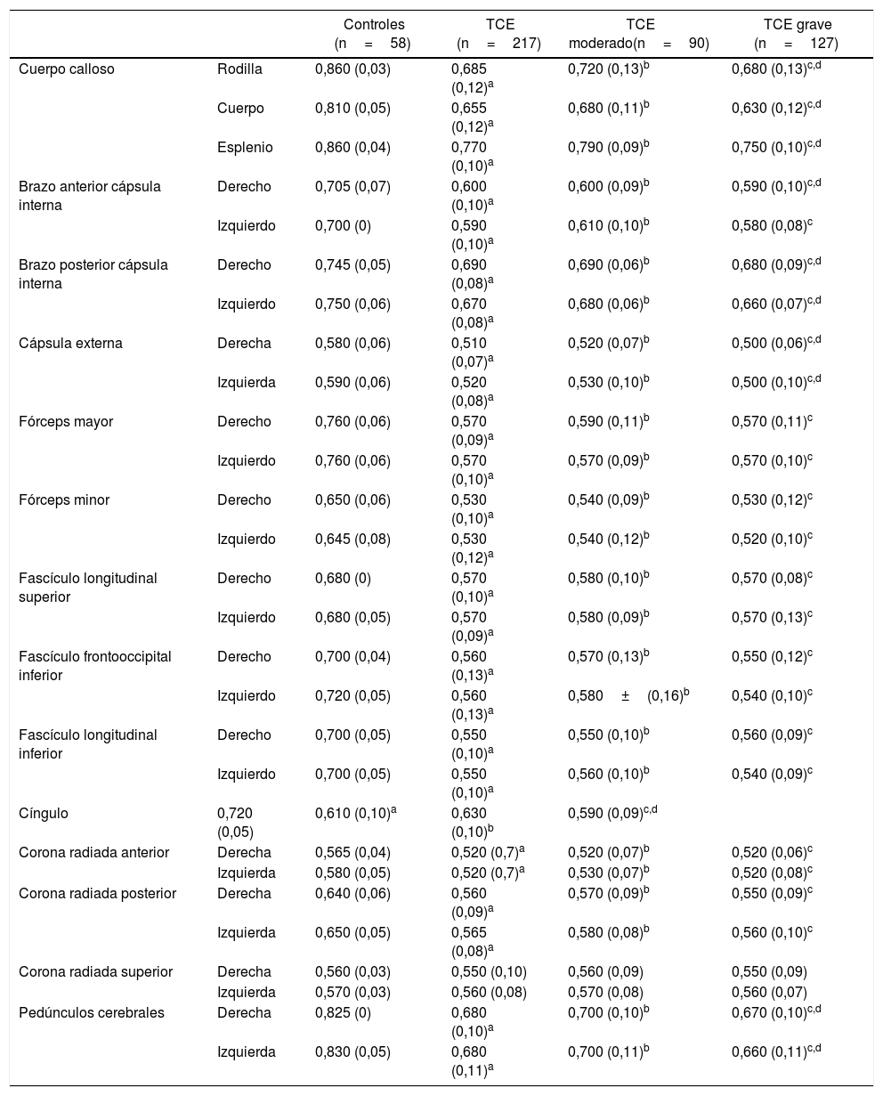
Materiales y métodosSe ha realizado RM-DTI a 217 pacientes con TCE moderado y grave en la fase subaguda precoz tras el TCE (mediana=19 días). El método de análisis elegido es por región de interés para calcular el valor medio de fractional anisotropy (FA) en 28 haces de sustancia blanca. Los valores obtenidos en los pacientes se han comparado con aquellos medidos en 58 sujetos sanos.
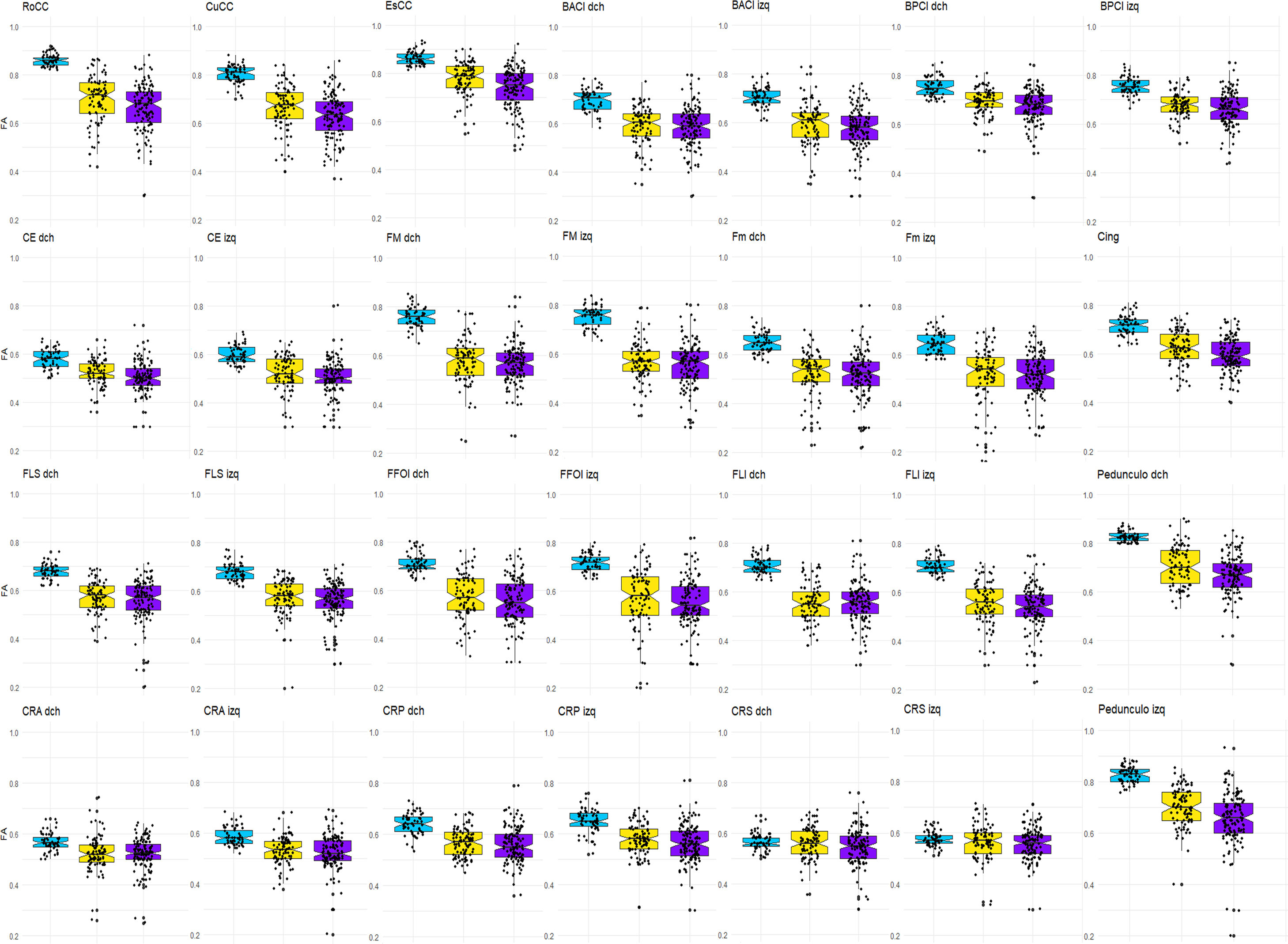
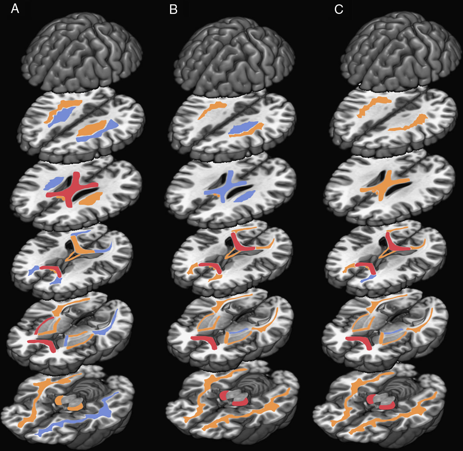
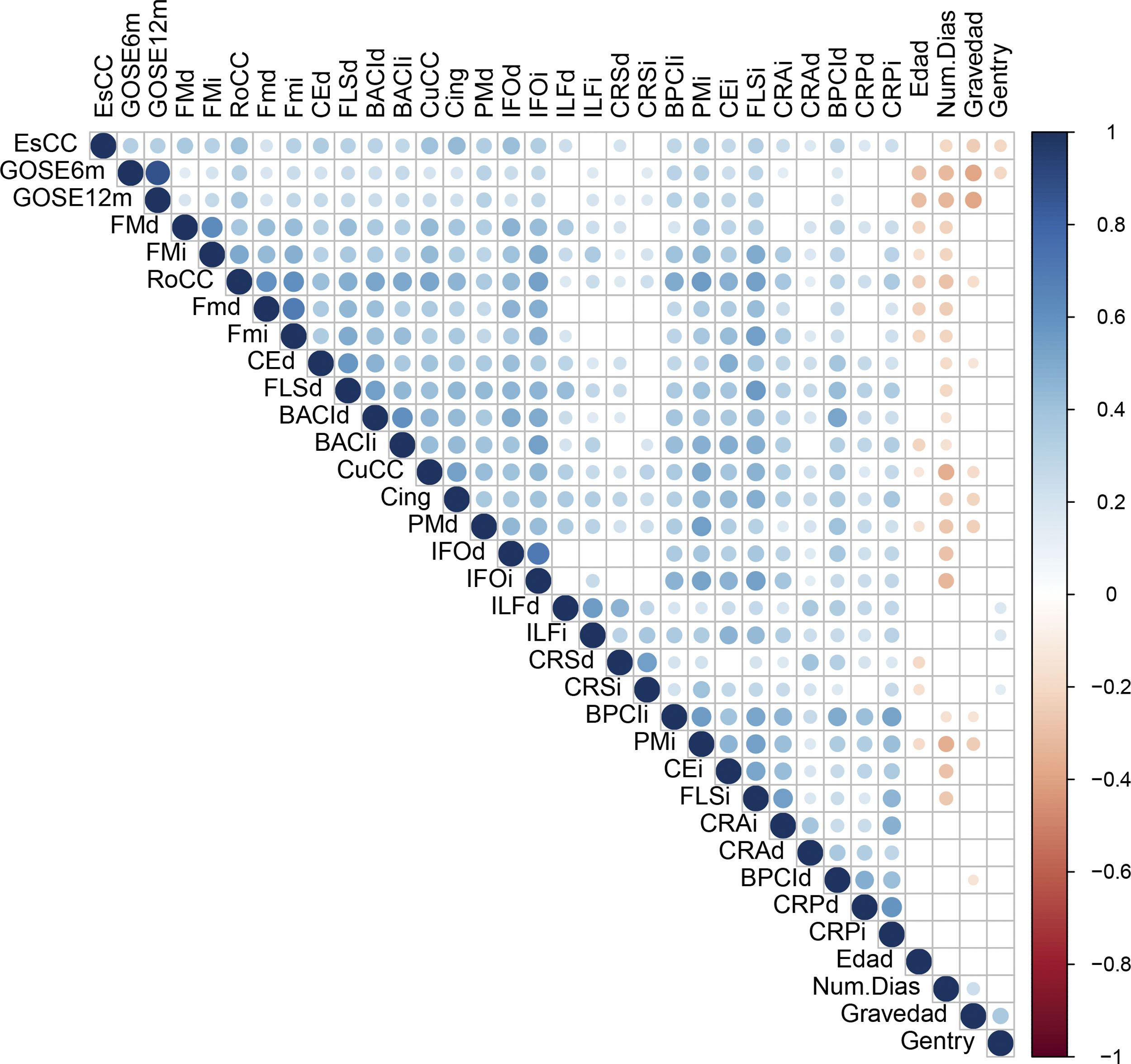
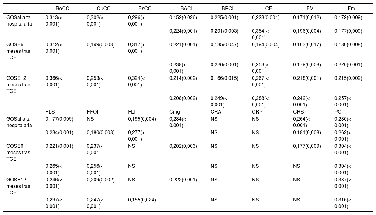
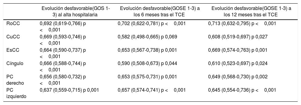
ResultadosLos resultados principales han sido que los pacientes, independientemente de la gravedad, demostraron valores de FA significativamente inferiores al grupo control en prácticamente todos los haces estudiados. Se detectó asociación entre el valor de FA y algunas variables clínicas de interés. Adicionalmente, los valores de FA de las tres porciones del cuerpo calloso, cíngulo y pedúnculos cerebrales se correlacionaron con la evolución del paciente evaluada a los 6 y 12 meses tras el TCE.
ConclusionesEl DTI es una herramienta útil para caracterizar la LAT y la detección de la reducción de FA en la fase subaguda precoz se relaciona con evolución desfavorable de los pacientes a medio y largo plazo.
Traumatic axonal injury (TAI) contributes significantly to mortality and morbidity after traumatic brain injury (TBI). Its identification is still a diagnostic challenge because of the limitations of conventional imaging techniques to characterized it. Diffusion tensor imaging (DTI) can indirectly identify areas of damaged white matter integrity by detecting water molecule diffusion alterations. Our main objective is to characterize the TAI using DTI at the early subacute stage in our series of moderate to severe TBI patients and to evaluate if there is a relationship between the information provided by the DTI and patient's outcome.
Materials and methodsWe have obtained DTI data from 217 patients with moderate to severe TBI acquired at a median of 19 days after TBI, and patient DTI metrics were compared with data obtained from 58 age-matched healthy controls. Region of interest method was applied to obtain mean fractional anisotropy (FA) value in 28 white matter fiber bundles susceptible to TAI.
ResultsOur main results were that when we compared patients with controls, patients, regardless of TBI severity, showed significantly reduced mean FA in almost all region of interest measured. We found statistically significant correlation between FA metrics and some clinical characteristics. Additionally, the FA values of the three portions of Corpus callosum, Cingulum and cerebral peduncles measured at the early subacute stage were highly associated with outcome assessed at hospital discharge and at 6 and 12 months after TBI.
ConclusionsWe conclude that DTI is a useful tool to characterize TAI and the detection of FA reduction in the subacute stage after TBI is associated with long-term unfavorable outcome.
Article
![]()
If it is the first time you have accessed you can obtain your credentials by contacting Elsevier Spain in suscripciones@elsevier.com or by calling our Customer Service at902 88 87 40 if you are calling from Spain or at +34 932 418 800 (from 9 to 18h., GMT + 1) if you are calling outside of Spain.
If you already have your login data, please click here .
If you have forgotten your password you can you can recover it by clicking here and selecting the option ¿I have forgotten my password¿.


