


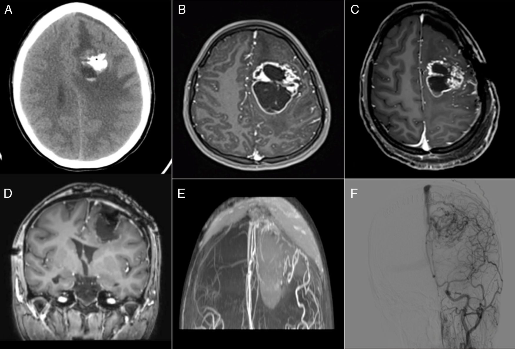
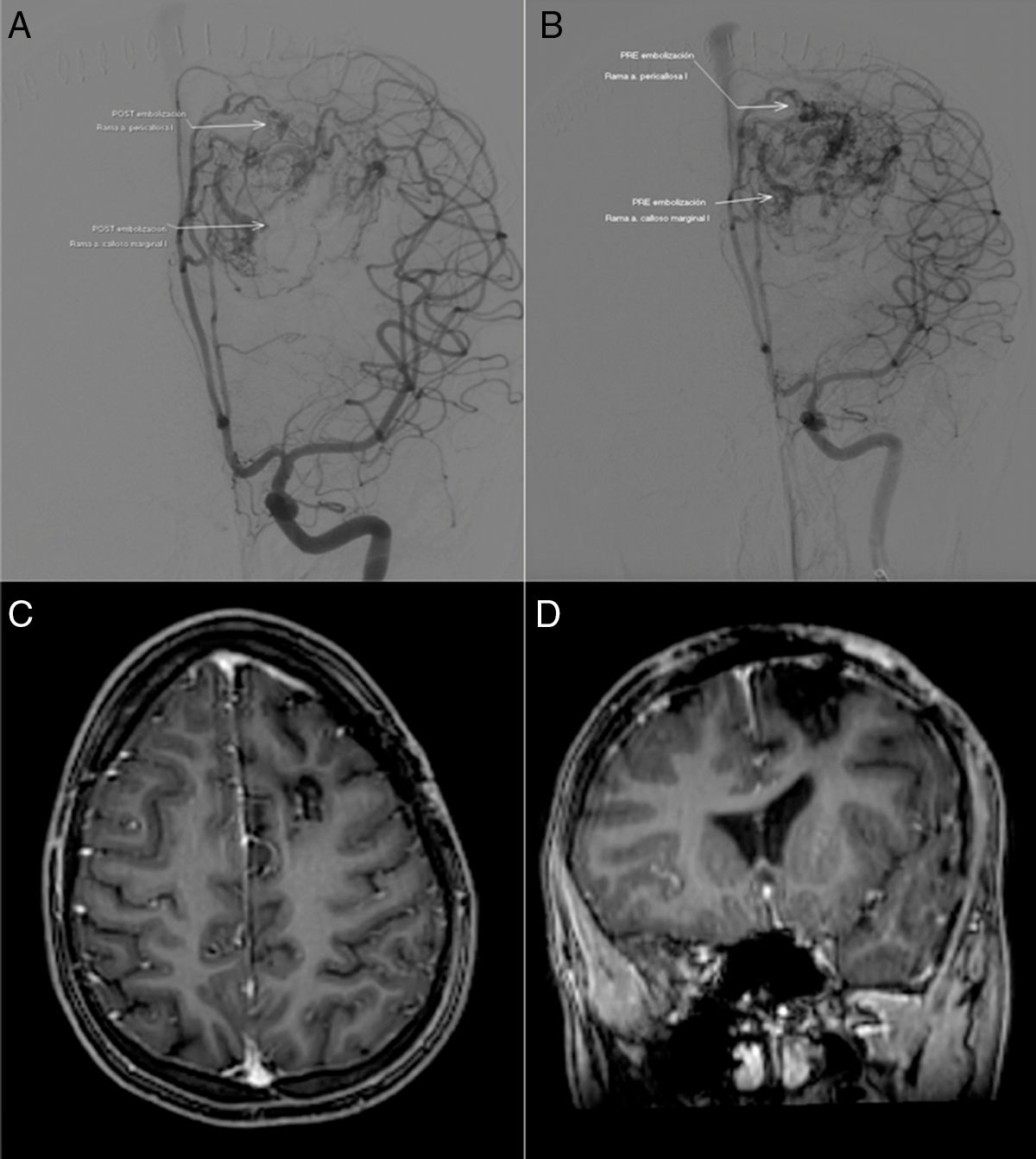
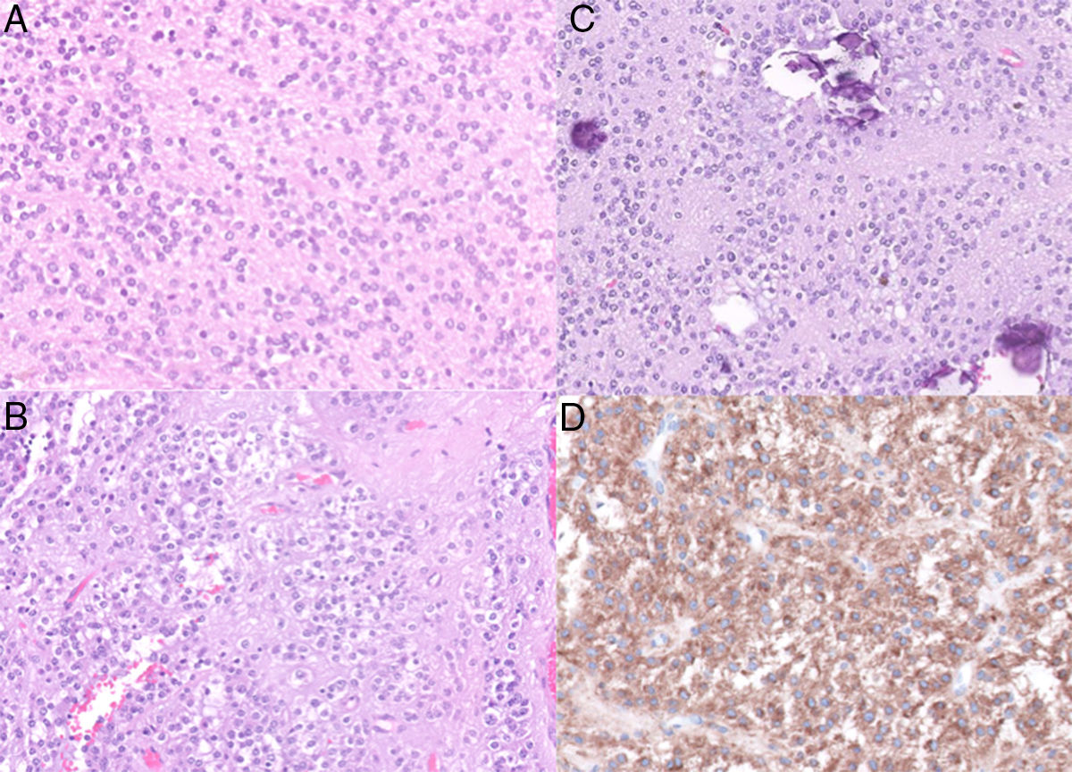
Una mujer de 13 años de edad presenta clínica de cefalea de 15 días de evolución y solo edema de papila bilateral en la exploración. El estudio inicial de tomografía computarizada y RM mostró una gran masa multiquística frontal izquierda con calcificación rodeada de edema periférico, sangrado intralesional subagudo y múltiples importantes vasos asociados. Se interviene en otro centro, encontrando cavidad con hematoma subagudo que se evacua con múltiples vasos y venas arteriolizadas. Ante la sospecha de malformación arteriovenosa (MAV) a pesar de los hallazgos de la neuroimagen realizada previamente, se deriva a nuestro centro para seguir tratamiento. Realizamos arteriografía, angio-RM y RM con secuencias avanzadas que muestran masa intraaxial hipervascularizada que se emboliza previo a la interviene quirúrgica definitiva con resultado anatomopatológico de neurocitoma extraventricular (NEV). Los NEV son lesiones extremadamente raras que no se han descrito previamente en la literatura como lesiones hipervascularizadas que en nuestro caso requirió la realización de angiografía y embolización previa para su correcto diagnóstico y adecuado manejo.
A 13-year-old female arrived at the Emergency Department with a two-week history of headache, and bilateral papilloedema on examination. The initial study with CT and MRI showed a large multicystic left frontal mass with calcification surrounded by peripheral oedema, subacute intralesional bleeding and association of multiple large vessels. She was initially operated on in another centre where a subacute haematoma was found, evacuating to multiple vessels and arteriolised veins. Despite the earlier neuroimaging findings, arteriovenous malformation (AVM) was suspected, so she was referred to our centre for further treatment. We performed angiography, MR angiography and MRI with advanced sequences, diagnosing a highly vascularised intra-axial tumour which was embolised. The patient was then definitively operated on, with the resulting finding of extraventricular neurocytoma (EVN). EVN are extremely rare lesions, not previously described in the literature as hypervascularised lesions, which in our case required prior angiography and embolisation for proper diagnosis and adequate management.
Article
![]()
If it is the first time you have accessed you can obtain your credentials by contacting Elsevier Spain in suscripciones@elsevier.com or by calling our Customer Service at902 88 87 40 if you are calling from Spain or at +34 932 418 800 (from 9 to 18h., GMT + 1) if you are calling outside of Spain.
If you already have your login data, please click here .
If you have forgotten your password you can you can recover it by clicking here and selecting the option ¿I have forgotten my password¿.


