C0239 - LIPOMATOUS EPENDYMOMAS OF THE POSTERIOR FOSSA. A VERY INFREQUENT AND LITTLE-KNOWN SUBTYPE
1Hospital Clínico San Carlos, Madrid, España. 2Hospital Son Espases, Mallorca, España. 3Hospital Puerta de Hierro, Madrid, España.
Objectives: To present a very infrequent subtype of ependymoma in the posterior fossa.
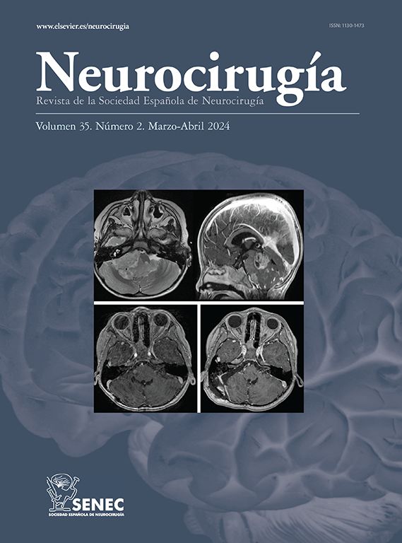
Methods: A 71-year-old woman with headache and gait instability, no cranial nerve alteration. No relevant personal history. CT showed Lesion in posterior fossa of heterogeneous appearance, hyperdense and calcifications in its interior. MRI Intraventricular infratentorial solid lesion. It measures approximately 5.0 × 4.0 × 3.0 cm (CC × T × AP), with the presence of edema/infiltration of the medial portion of the left cerebellar hemisphere and the ipsilateral cerebellar peduncle. It occupies extensively the IV ventricle and extends through Magendie and both Luschkas to the posterior region of the foramen magnum.
Results: Histology: neoplastic cell proliferation of glial strain of low cytological aggressiveness, conformation of few gliovascular pseudorosettes and rare ependymal rosettes. The cells have vacuolated broad cytoplasm with eccentric nuclei, reminiscent of adipocytes and cells in a signet-ring. Absence of mitosis, necrosis and vascular proliferation. The stains for mucins are negative. The immunohistochemical study reveals that the neoplastic cells widely express PFGA, CD99, S100, CKAE1AE3, CK7, NSE, COX2, Vimentin and RCC and are negative for EMA and CK20. Although the neoplasm is of unusual morphology, the typical histological findings, although scarce, together with the immunophenotype of the neoplastic cells and their intraventricular location confirm the diagnosis of lipomatous ependymoma (grade II).
Conclusions: The lipomatous histopathological pattern, whose cells resemble adipocytes and signet-ring cells, is not conventional in ependymomas and its content is under debate. In these, a ballooning of the cellular body is observed by a possible accumulation of metabolic products or hyperplastic organelles that move the nucleus to the periphery. It is believed that vacuoles are the result of microrosettes and/or an inflamed and degenerated cytoplasm. it is important not to confuse this variant with more common lesions, such as metastatic adenocarcinoma especially those of the breast, intestine or renal.


