Journal Information
Vol. 26. Issue 4.
Pages 200-204 (July - August 2015)
Share
Share
Download PDF
More article options
Vol. 26. Issue 4.
Pages 200-204 (July - August 2015)
Caso clínico
Malformación arteriovenosa intracraneal múltiple
Multiple intracranial arteriovenous malformation
Miguel Gelabert-Gonzáleza,
Corresponding author
miguel.gelabert.gonzalez@sergas.es

Autor para correspondencia.
, José María Santin-Amoa, Paula Román-Penaa, Fernando Vázquez Herrerob
a Servicio de Neurocirugía, Hospital Clínico Universitario de Santiago de Compostela, Departamento de Cirugía, Universidad de Santiago de Compostela, Santiago de Compostela, La Coruña, España
b Servicio de Radiología, Hospital Clínico Universitario de Santiago de Compostela, Santiago de Compostela, La Coruña, España
Article information
Abstract
Full Text
Bibliography
Download PDF
Statistics
Figures (3)
Show moreShow less
Resumen

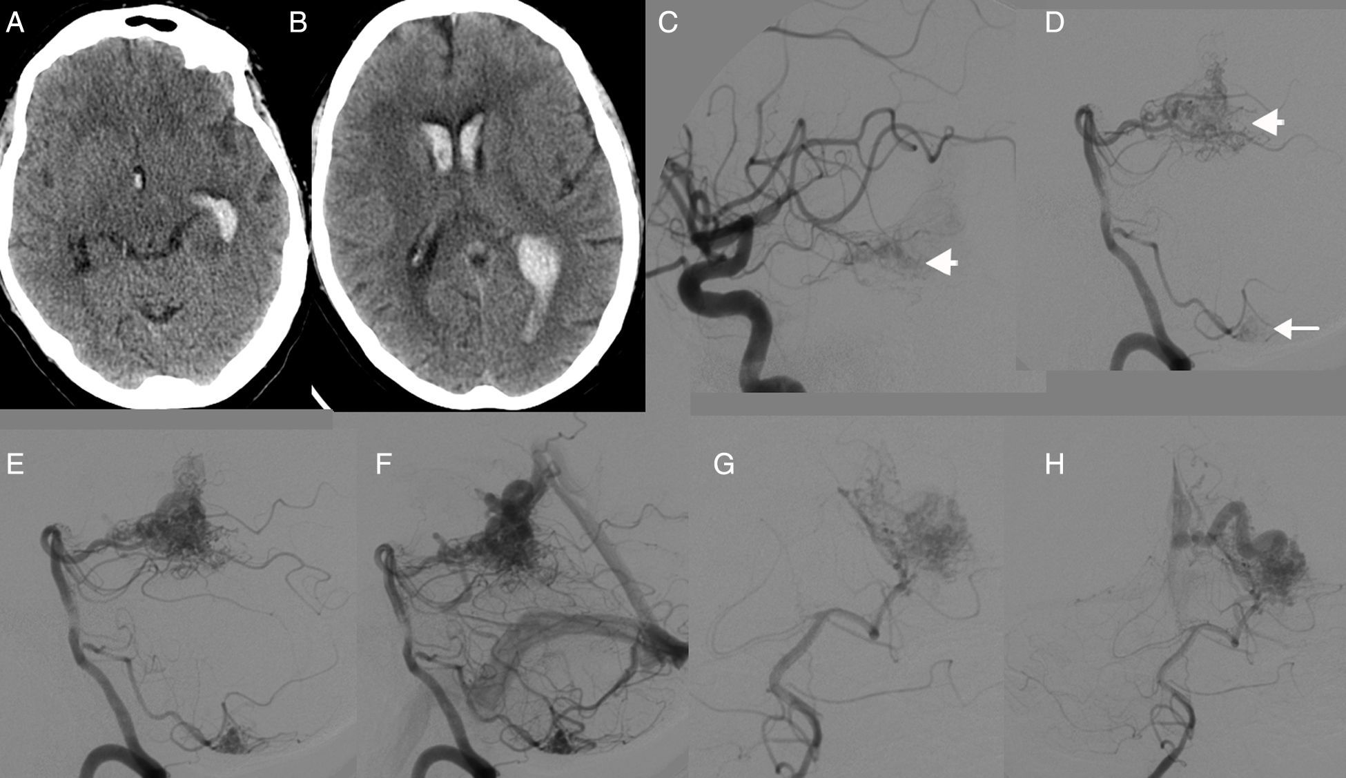
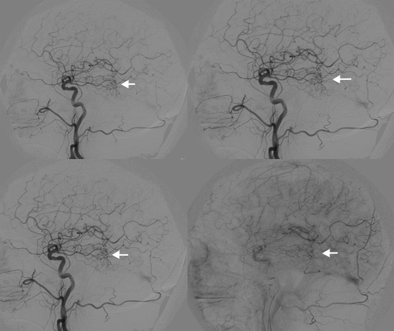
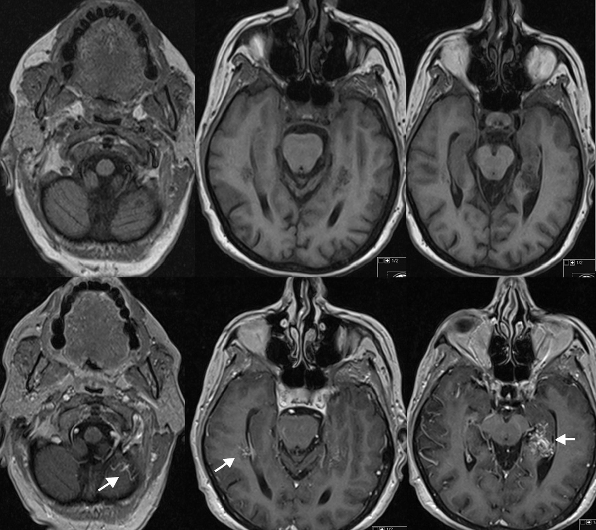
Las malformaciones arteriovenosas (MAV) cerebrales múltiples son poco frecuentes y la mayoría de las publicaciones existentes presentan casos clínicos aislados. Su incidencia en grandes series de MAV oscila entre el 0,3 y 9%, y en la mayoría de casos aparecen asociadas con otras anomalías vasculares del cerebro u otros tejidos. Presentamos el caso clínico de una mujer de 62 años que sufrió una hemorragia parenquimatosa temporal izquierda y que en los estudios neuro-radiológicos se evidenciaron 3 MAV localizadas en: lóbulo temporal izquierdo, hemisferio cerebeloso izquierdo y lóbulo temporal derecho. Las lesiones fueron tratadas con radiocirugía.

Palabras clave:
Angiografía cerebral
Malformación arteriovenosa múltiple
Hemorragia parenquimatosa
Radiocirugía
Abstract

Multiple cerebral arteriovenous malformations (AVMs) are thought to be exceedingly rare lesions and have usually been reported as single cases. The incidence of multiple cerebral AVMs in major series ranges from 0.3% to 9% and, in the majority of cases, these malformations are associated with other vascular anomalies of the brain or soft tissues. We report a 62-year-old woman that presented with a left temporal haemorrhage. Angiography showed 3 AVMs located in the left temporal lobe, left cerebellar hemisphere and right temporal lobe. The lesions were treated with radiosurgery.

Keywords:
Angiography
Brain haemorrhage
Multiple arteriovenous malformations
Radiosurgery

Article

These are the options to access the full texts of the publication Neurocirugía (English edition)
Member
Member of the Sociedad Española de Neurocirugía

If it is the first time you have accessed you can obtain your credentials by contacting Elsevier Spain in suscripciones@elsevier.com or by calling our Customer Service at902 88 87 40 if you are calling from Spain or at +34 932 418 800 (from 9 to 18h., GMT + 1) if you are calling outside of Spain.

If you already have your login data, please click here .

If you have forgotten your password you can you can recover it by clicking here and selecting the option ¿I have forgotten my password¿.

Subscriber
Subscriber

If you already have your login data, please click here .

If you have forgotten your password you can you can recover it by clicking here and selecting the option “I have forgotten my password”
Subscribe
Subscribe to

Neurocirugía (English edition)

Purchase
Purchase article

Purchasing article the PDF version will be downloaded

Price 19.34 €

Purchase now
Contact
Phone for subscriptions and reporting of errors
From Monday to Friday from 9 a.m. to 6 p.m. (GMT + 1) except for the months of July and August which will be from 9 a.m. to 3 p.m.
Calls from Spain
932 415 960
Calls from outside Spain
+34 932 415 960
Email
Idiomas
Neurocirugía (English edition)
es en

¿Es usted profesional sanitario apto para prescribir o dispensar medicamentos?

Are you a health professional able to prescribe or dispense drugs?