


Conocer la anatomía microquirúrgica del complejo venoso petroso superior (CVPS).
Material y métodosSe realizó estudio descriptivo y prospectivo. Se utilizaron 6 especímenes (12 lados) inyectados. Se estudió la anatomía microquirúrgica del CVPS en los encéfalos, mediante un abordaje retrosigmoideo y transpetroso anterior. Se utilizó instrumental neuroquirúrgico, endoscopio rígido de 0 grados, microscopio quirúrgico OPMI-1 con magnificación 6× a 20×. Se analizó el patrón de drenaje hacia el seno petroso superior, la formación de las venas tributarias, la relación con el nervio trigémino y las variantes del CVPS.
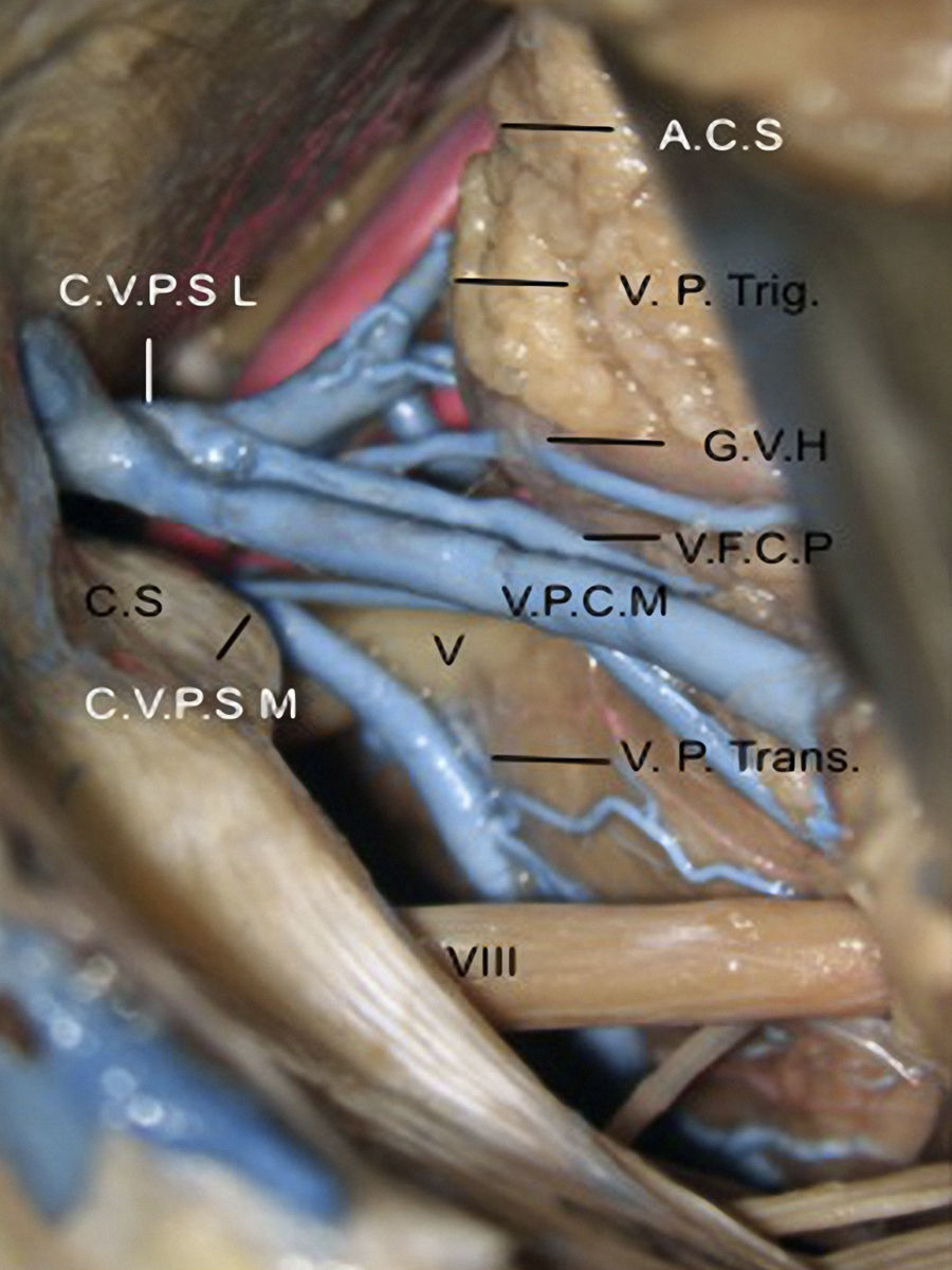
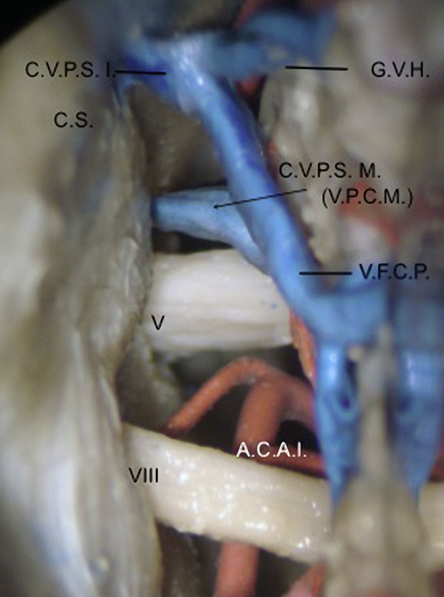
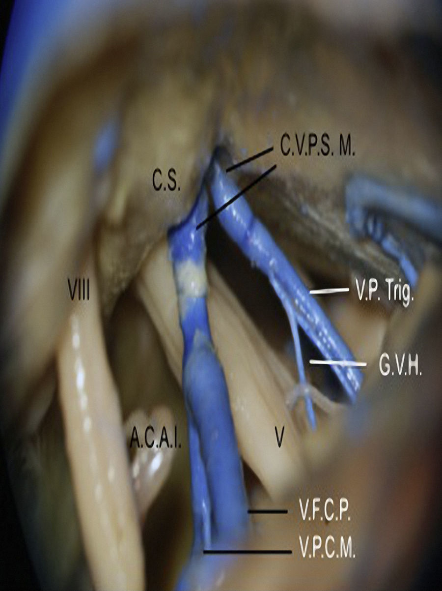
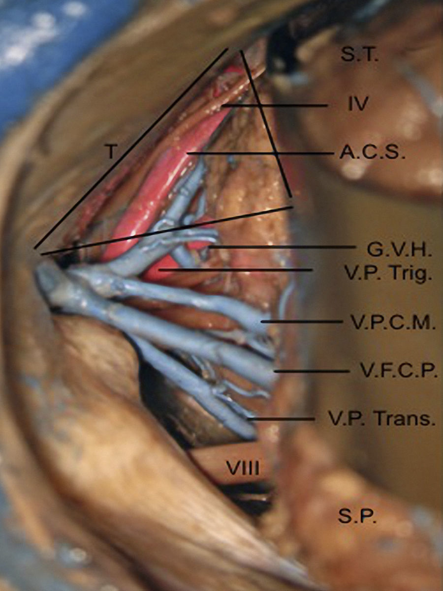
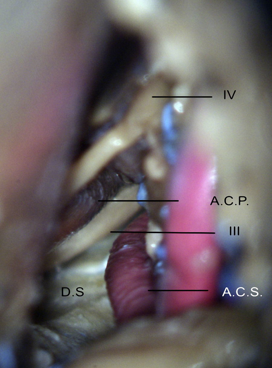
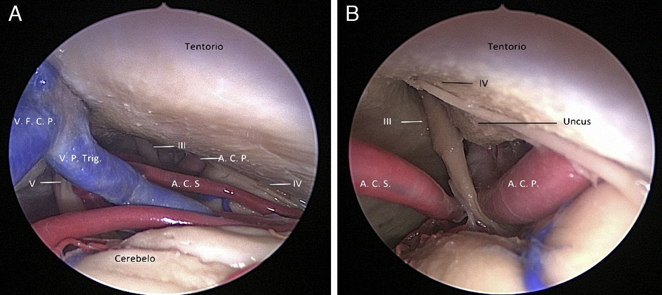
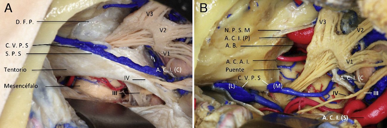
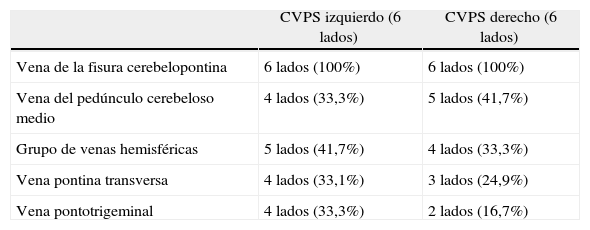
ResultadosEl CVPS se encontró en todos los lados, la vena tributaria que se encontró en el 100% de los lados fue la vena de la fisura cerebelopontina. El patrón de drenaje del CVPS fue dividido en relación a la cresta suprameatal en: lateral, medial y en un punto intermedio. Se encontró el CVPS simple, formado por un tronco con sus tributarias, en 8 lados y duplicado en 4 lados. En el estudio se observó un triángulo formado por el tentorio, el CVPS y parte de la cara petrosa y tentorial del cerebelo con bordes y contenidos bien definidos, el cual se llamó triángulo petroso-tentorial.
ConclusiónEs necesario entender la anatomía microquirúrgica del CVPS. Así, proponemos el triángulo petroso-tentorial como corredor neuroquirúrgico para el manejo de lesiones del ángulo pontocerebeloso a la región petroclival superior.
To study the microsurgical anatomy of the superior petrosal venous complex (SPVC).
Material and methodsWe conducted a descriptive and prospective study. Six injected specimens were used (12 sides). The microsurgical anatomy of the SPVC was studied by means of an anterior, retrosigmoid and transpetrosal approach. Neurosurgical equipment, 0-degree rigid endoscopy and OPMI-1 surgical microscope with 6× to 20× magnification were all used in this study. The venous drainage pattern toward the superior petrosal sinus was analysed, as were the formation of tributary veins, the relationship with the trigeminal nerve and the anatomical variants of SPVC.
ResultsThe SPVC was present in all cases. A tributary, cerebellopontine fissure vein was identified in 100% of cases. The venous drainage pattern of the SPVC was divided into medial, intermediate and lateral with respect to the suprameatal crest. The SPVC was simple in 8 sides and duplicate in 4 sides. A triangle formed by the tentorium, the SPVC and part of the tentorial and petrosal surface of the cerebellum was also observed in the study. This triangle was called the petrosal-tentorial triangle.
ConclusionsIt is important to understand the microsurgical anatomy of the SPVC. Therefore, we propose the petrosal-tentorial triangle as a neurosurgical route for the management of pathologies from the cerebellopontine angle to the superior petroclival region.
Article
![]()
If it is the first time you have accessed you can obtain your credentials by contacting Elsevier Spain in suscripciones@elsevier.com or by calling our Customer Service at902 88 87 40 if you are calling from Spain or at +34 932 418 800 (from 9 to 18h., GMT + 1) if you are calling outside of Spain.
If you already have your login data, please click here .
If you have forgotten your password you can you can recover it by clicking here and selecting the option ¿I have forgotten my password¿.


