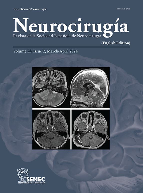
En la extirpación completa del neurinoma acústico por vía suboccipital retrosigmoidea, es obligada la apertura de la pared posterior del conducto auditivo interno (CAÍ). Por lo tanto, uno de los pasos clave en el abordaje quirúrgico transmeatal es el fresado del CAÍ. Sin embargo, no existen claras referencias anatómicas intraoperatorias para la identificación de estructuras tales como los canales semicirculares, el golfo de la vena yugular o las celdas aéreas. Las variaciones anatómicas individuales y las producidas por el propio tumor, obligan en cada caso a una correcta planificación preoperatoria, si queremos evitar complicaciones secundarias a su lesión yatrógena (cófosis, licuorrea, hemorragia y embolismo aéreo).
ObjetivoSe expone la experiencia del primer autor firmante (EU) en el fresado del CAÍ con especial referencia a la topografía anatómica y límites quirúrgicos en el abordaje suboccipital retrosigmoideo a la porción intracanalicular del neurinoma acústico.
Material y métodosEste trabajo está basado en datos anatómicos obtenidos del fresado de huesos temporales normales extraídos de material autópsico junto a nuestra experiencia sobre 20 pacientes intervenidos de neurinoma acústico siguiendo la técnica y protocolo de Samii.
ResultadosNo hemos intervenido por esta vía ningún tumor puramente intracanalicular. 2 casos han sido de grado II (hasta 20mm de diámetro), 12 de grado III y 6 casos de grado IV. En ningún caso se ha llegado a fresar tanto como para visualizar el fondo del CAÍ, lo que se confirmó con el TAC postoperatorio; a pesar de ello en 17 casos se ha considerado la extirpación como completa. No ha existido mortalidad y no hemos tenido complicaciones mayores atribuidas al fresado del CAÍ, como licuorrea o embolismo aéreo. No podemos asegurar que la hipoacusia o la cófosis postoperatoria, que han sido la regla excepto en un caso de grado II, haya sido causada por lesión nerviosa, laberíntica o isquémica.
ConclusionesEn nuestro material no ha sido posible la exposición completa del fondo del CAÍ por vía retrosigmoidea sin lesionar alguna estructura laberíntica. Las zonas de mayor riesgo de complicaciones secundarias al fresado son la pared inferior y el fondo del CAL La extensión medial de la craniectomía suboccipital facilita al fresado y a la exposición tumoral intrameatal. No existen referencias intraoperatorias para localizar las estructuras petrosas durante el fresado del CAÍ excepto la propia experiencia del cirujano.
To completely remove the intracanalicular portion of the acoustic neuroma through the retrosigmoid approach, we must open the posterior wall of the internal auditory canal (IAC). Therefore, drilling the IAC is one of the key steps we need to take in the transmeatal surgical approach. Nevertheless, there are no clear anatomical landmarks to identify structures such as the semicircular cañáis, the jugular bulb or air cells. The individual anatomical variations and those caused by the tumour itself make preoperative evaluation essential if we wish to avoid complications such as deafness, cerebrospinal fluid leakage, bleeding and air embolism.
ObjectiveWe describe here the personal experience of the sénior author (EU) in drilling the posterior wall of the IAC, with special reference to the anatomical landmarks and surgical limits in the suboccipital approach to the intracanalicular portion of the acoustic neuromas.
Material and methodsThis work is based on anatomical data obtained from drilling human temporal bones obtained from cadavers, along with our experience with 20 patients who were operated on for acoustic neuroma using Samii's technique.
ResultsWe did not opérate on any purely intracanalicular neurinomas using this approach. Two tumors were grade II (up to 20mm in diameter), 12 were grade III and 6 were grade IV. We did not drill far enough in any of these cases to be able to see the fundus of the IAC, which was conflrmed by postoperative CT. Despite this, the tumour was considered to be completely removed in 17 cases. There was no mortality and we had no major complications as a result of drilling the IAC such as cerebrospinal fluid leakage or air embolism. We cannot guarantee that hearing loss or postoperative deafness, which were the norm except in one case of grade II, were caused by nervous, ischemic or labyrinthine lesions.
ConclusionsIn our material it was not possible to completely expose the IAC fundus using a retrosigmoid approach without injury to the labyrinth. The áreas in which the risk of secondary complications is greatest when drilling are the inferior wall and the IAC fundus. The medial extensión of the suboccipital craniotomy makes drilling and intrameatal tumour exposure easier. There are no intraoperative landmarks to lócate the petrous structures while drilling the IAC except for those provided by the surgeon's own experience
Article

If it is the first time you have accessed you can obtain your credentials by contacting Elsevier Spain in suscripciones@elsevier.com or by calling our Customer Service at902 88 87 40 if you are calling from Spain or at +34 932 418 800 (from 9 to 18h., GMT + 1) if you are calling outside of Spain.
If you already have your login data, please click here .
If you have forgotten your password you can you can recover it by clicking here and selecting the option ¿I have forgotten my password¿.


